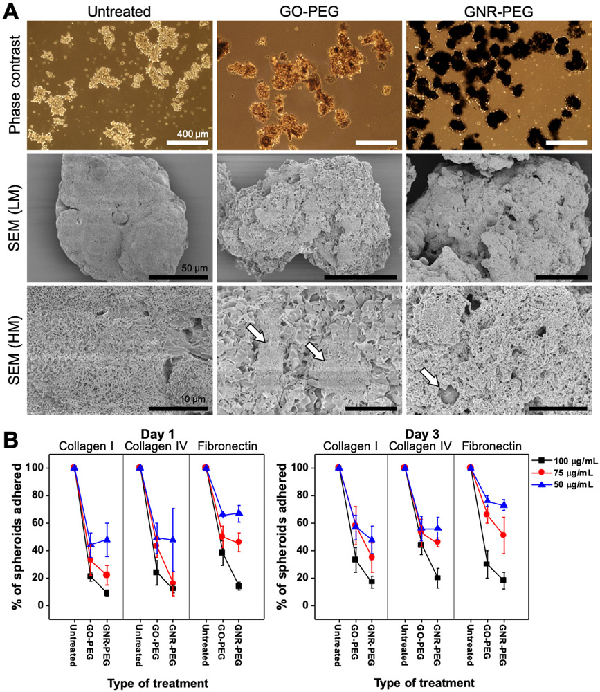
Figure 2.
GO-PEG and GNR-PEG disrupt adhesion of SKOV-3 spheroids to ECM proteins abundant in the mesothelial layer. (A) Representative phase contrast and SEM images of SKOV-3 spheroids (LM; low magnification, HM; high magnification). Scale bars indicate 400 μm in phase contrast images and 50 μm (LM) or 10 μm (HM) in SEM images. The white arrows in SEM (HM) images indicate exposed cell surfaces. (B) Percentage of total spheroids that have adhered after 1 or 3 days of incubation on collagen I, collagen IV, or fibronectin.

