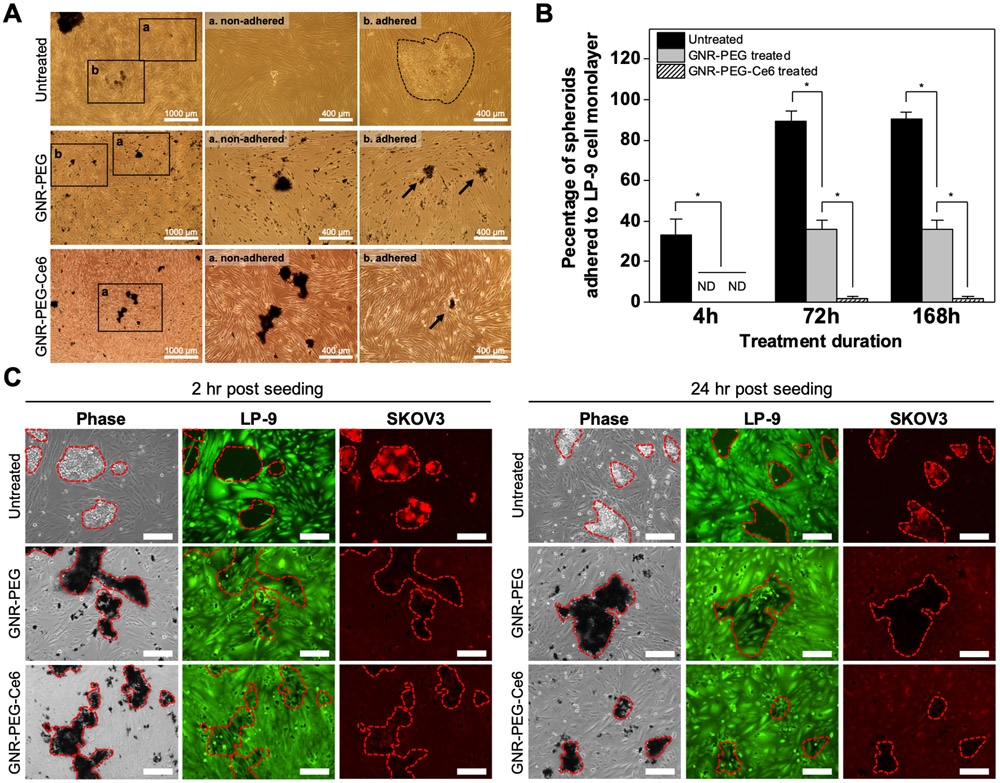
Figure 4.
GNR-PEG-Ce6 disrupts mesothelial adhesion of SKOV-3 spheroids and subsequent mesothelial clearance. (A) Representative phase contrast images of adhered and non-adhered spheroids in each condition after 72 h of incubation. (B) Percentage of total spheroids that have adhered to the LP-9 mesothelial cell layer after 4, 72, or 168 h of incubation (*P < 0.05, ND indicates not detected). (C) Representative phase contrast images of SKOV-3 spheroids breaching the underlying LP-9 monolayer and corresponding fluorescence images of the LP-9 monolayer (green) and SKOV-3 spheroids (red) at 2 or 24 h after spheroid seeding. Scale bars indicate 200 μm.

