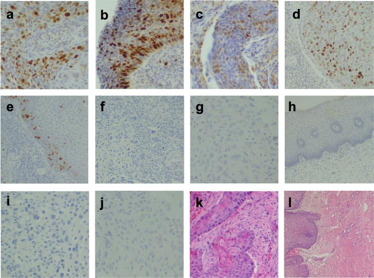
Fig 1. Monographs.
Nucleus and cytoplasm were positively stained for (a-b) p16 at 200x for cancerous and precancerous cervical lesion respectively. Nucleus positively stained for (c-d) TOP2A at 200x for cancerous and precancerous cervical lesion respectively, and (e) TOP2A at 40x for a benign cervical lesion. Nucleus and cytoplasm negatively stained for (f-g) p16 at 100x and 200x for cancerous and precancerous cervical lesion respectively, and (h) p16 at 200x for a benign cervical lesion. Nucleus negatively stained for (i-j) TOP2A at 200x for cancerous and precancerous cervical lesion respectively; (k-l) H&E staining at 100x and 40x for squamous cell carcinoma and normal cervix respectively.

