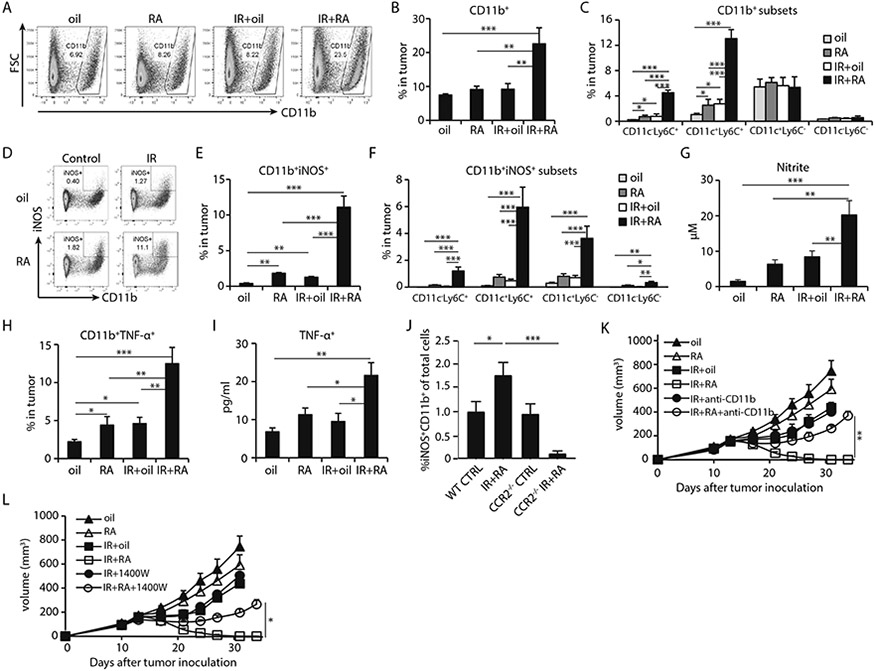
Fig. 2. Combining local ablative IR with RA induces iNOS and TNF-α producing inflammatory macrophages.

MC38 tumors were treated as indicated and harvested 4 days post start of the IR/RA treatments for analysis in A-J.
(A) The gating strategy of iNOS+ myeloid cells.
(B) Frequency of CD11b+ myeloid cells in total cells of tumors.
(C) Percentage of major subsets of CD11b+ cells in tumors received IR/RA treatment.
(D) Gating strategy of iNOS+ inflammatory macrophages (Inf-MACs).
(E) Percentage of iNOS+ inflammatory macrophages in total cells of tumors.
(F) Percentage of subsets of iNOS+ Inf-MACs in terms of CD11c and Ly6C marker in total cells.
(G) Concentration of iNOS product nitrite in tumor homogenate. Treated tumors were harvested on day 4 post treatment and tumor fragments were cultured in vitro. Nitrite level in culture media was measured after 2 hours.
(H) TNF-α+ CD11b+ population in tumor receiving indicated treatment.
(I) Concentration of TNF-α in tumor homogenate at 4 days after start of the indicated treatments.
(J) Percentage of Inf-MACs in tumors grown in WT or CCR2−/− mice, 4 days after start of IR and RA treatment.
(K) Tumor growth curve of MC38 tumors during treatments while CD11b+ cell recruitment was blocked.
(L) Tumor growth curve of MC38 tumors during treatments while iNOS inhibitor (1400W) was administered.
*, p<0.05; **, p<0.01; ***, p<0.001. Experiments were conducted 3 times with 3-5 mice in each group. Data in K, L are presented as mean ± SEM, the rest of the data are mean ± SD. Results from representative experiments are shown.
