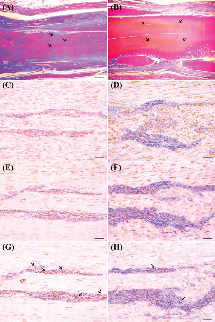
FIGURE 6.

Tissues sections obtained from sheep CCT, after induction of collagenase type I iatrogenic lesions, injected or not with SFV cells, and sampled 8 weeks after therapy. Tendons stained with Herovici polychrome stain, highlight the longitudinal orientation of collagen fibers after transplantation of SFV cells, without necrotic and/or edematous aspect (A, scale bar = 13.3 μm); note that, in central area of the treated tendon, not completely mature, and longitudinal configured collagen fibers, identified as blue stained fibers admixed with red ones. In the control tendon (B, scale bar =13.3 μm), Herovici stain shows the presence of mature and longitudinally oriented collagen fibers (ie, predominantly strong Herovici red stained fibers in tendon sections) at the periphery of the collagenase injected area (arrows). In the central part of CCT, lesion (stained in yellowish—orange) is surrounded by completely mature, and fingerprint configured collagen fibers, identified as red stained fibers without pre‐collagen admixed blue fibers. Immunohistochemistry stains for collagen type I (CI), and collagen type III (CIII) show a high expression of CI (C, scale bar = 250 μm) and a very low expression of CIII (E, scale bar = 250 μm) in SVF cells—treated tendon. On the contrary, an opposite trend is observed in untreated control tendon in which a low expression of CI (D, scale bar = 250 μm), and high expression of CIII (F, scale bar = 250 μm) are observed. IHC stains for FVIIIRa reveal a large number of small and medium sized positive—micro‐vessels, in SVF cells—treated CCT (arrows) (G, scale bar = 250 μm). Although some rare endothelial cells are positive, a substantial absence of neo‐angiogenesis is observed in the control tendon, where only the presence of some small capillaries is sporadically observed (H, scale bar = 250 μm)
