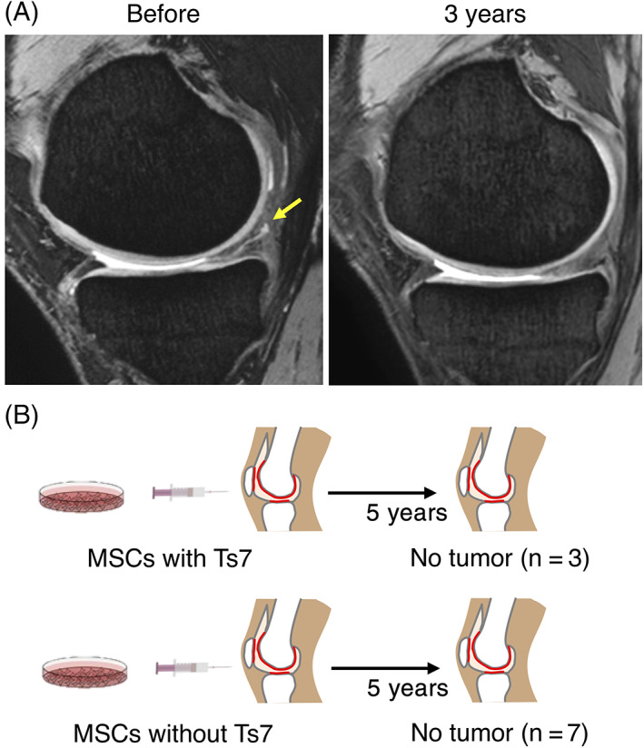
FIGURE 6.

Follow‐up of 10 patients for 5 years after mesenchymal stem cell (MSC) transplantation. A, Representative magnetic resonance imaging (MRI) of the meniscus before and 3 years after cell transplantation. Yellow arrow shows a meniscus degenerative tear. (B) Follow‐up results on tumorigenesis
