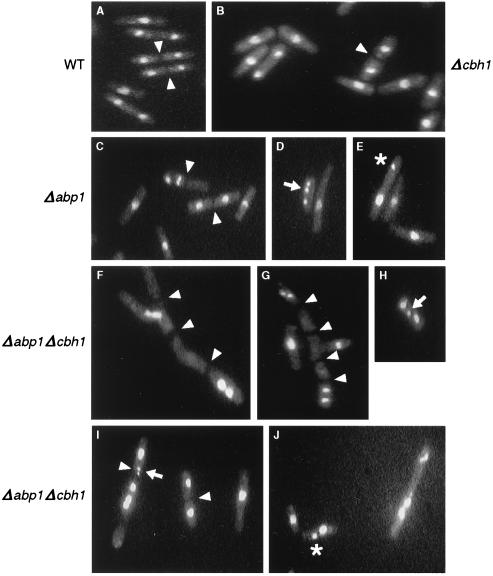
FIG. 5.
Chromosome missegregation and defective cytokinesis in a Δabp1 Δcbh1 strain. Ethanol-fixed, propidium iodide-stained cells were examined by fluorescence microscopy as described in footnote a to Table 3. Wild-type (A) and Δcbh1 (B) cells appeared uniformly normal, whereas Δabp1 (C to E) cells showed rare instances of missegregation. In contrast, Δabp1 Δcbh1 (F to J) cells showed frequent mistakes in chromosome segregation, as well as many cells with branched, multiseptated morphology (magnification, ×120). Cell septa (arrowheads), lagging chromosomes (arrows), and unequal distribution of chromatin masses (asterisks) are indicated. (See Tables 3 and 4 for a summary of the data). All cultures were grown at 32°C.

