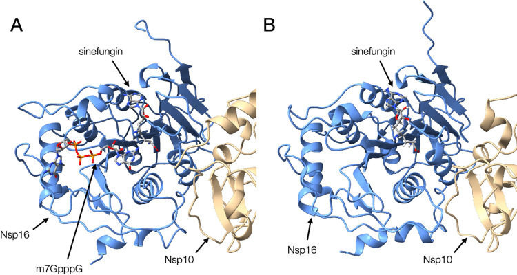
Figure 3 .
Comparison of structures of Nsp10-Nsp16 complex. (A) The homology model of SARS-CoV-2 Nsp10 (beige) and Nsp16 (blue) complex bound to Sinefungin and m7GpppA, which were based on the structure of SARS-CoV as the template in our previous study (YP_009725311_v02_1.pdb from BSM-Arc [85] entry BSM-ID BSM00015). (B) The crystal structure of the Nsp10-Nsp16 complex from SARS-CoV-2 bound to Sinefungin [PDB ID: 6yz1].

