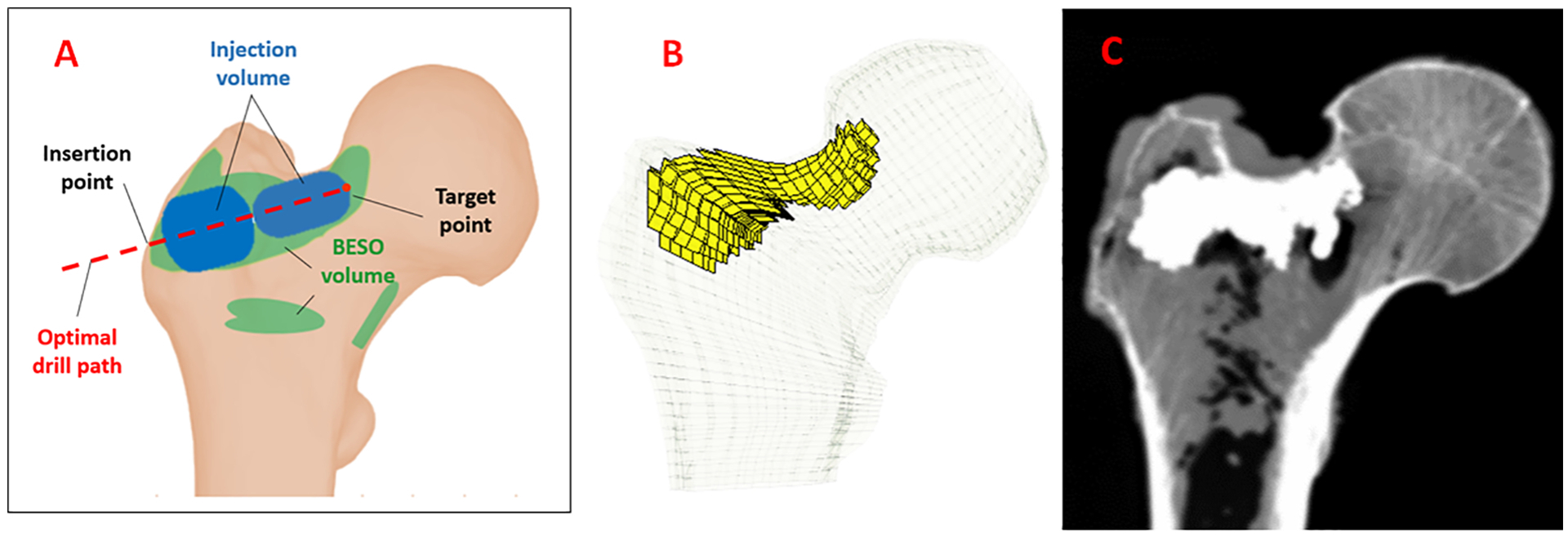
Figure 1 –

A) Schematic of a typical optimized injection pattern by FE using BESO method (green) and the injection blobs defined by the planning paradigm (Blue), B) FE mesh of the femur specimen with planned pattern of injection C) Example radiograph of the augmented bone (sample 1).
