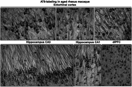
Figure 10.

Tau fibrillation (AT8 labeling) in aged rhesus macaque (31 years). Dense tau fibrillation is observed aggregating in entorhinal cortex layer II cell islands (a), extending into the deeper layers, including layer III (b) and layer V (c). The fibrillated tau‐labeling pattern is especially prominent in the perisomatic compartment and along apical and basilar dendrites (pretangles) of excitatory neurons, including parallel bundles of “twisting” apical dendrites in entorhinal cortex layer III and layer V, indicative of possible neurodegeneration. Prominent aggregated AT8 labeling is visualized in AD‐related vulnerable brain regions, including hippocampus pyramidal cell CA3 (d, e) and CA1 (f) subfields, and recurrent excitatory circuits in dlPFC layer III (g). Scale bars, 25 µm (a–c) and 30 µm (d–g). AD, Alzheimer's disease; dlPFC, dorsolateral prefrontal association cortex
