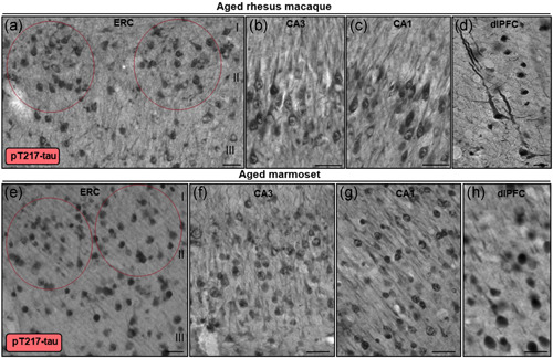
Figure 12.

Multiregional pT217‐tau labeling in aged rhesus macaque and marmoset. In aged rhesus macaque (30–31 years), stellate cell layer II islands (red ovals) in entorhinal cortex (a), hippocampus CA3 (b), hippocampus CA1 (c), and dlPFC layer III (d) are reactive against pT217‐tau. Immunoreactivity for pT217‐tau is visualized in the cytoplasm of the perisomatic compartment, along apical and basilar dendrites, and within the nucleoplasm. Note, in aged rhesus macaque dlPFC (30 years), pT217‐tau labeling reveals a highly fibrillated pattern, reflected in “twisting” apical dendrites, similar to AT8. In aged marmoset (12 years), stellate cell layer II islands (red ovals) in entorhinal cortex (e), hippocampus CA3 (f), hippocampus CA1 (g), and dlPFC layer III (h) are reactive against pT217‐tau. Immunoreactivity for pT217‐tau in aged marmosets is more diffuse compared to rhesus macaques, but observed prominently in the neuronal soma, and proximal and distal segments of apical dendrites. Scale bars, 25 µm (a–c, e–g), 10 µm (d), 20 µm (h)
