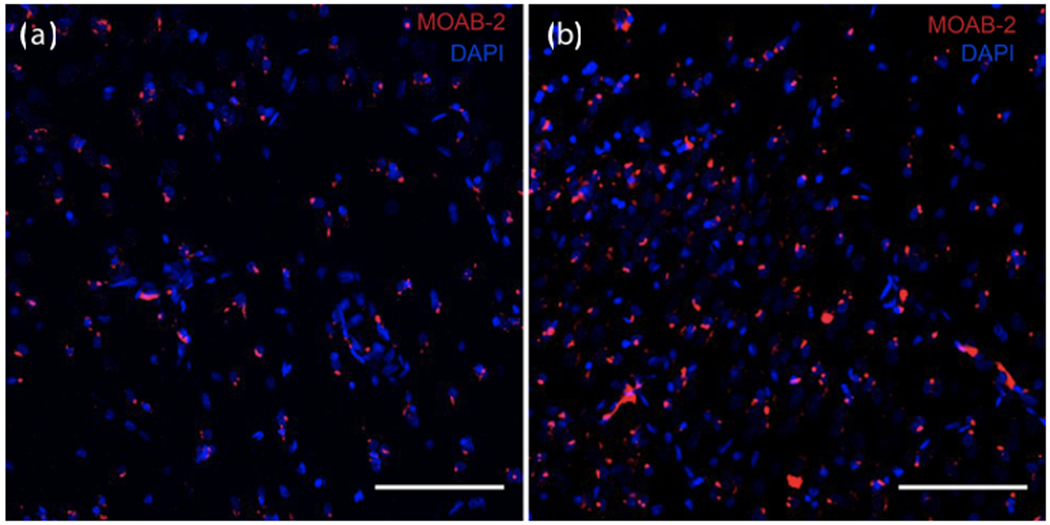
Figure 2. Amyloid deposits in hippocampus.

Varying degrees of amyloid deposits in CA1 of old marmosets. Amyloid-β peptide detected by MOAB-2 antibody (red); cell nuclei stained with DAPI (blue). Scale bar = 100 μm. Amyloid burden in two male marmosets: (a) low burden, 9 years old and (b) high burden 8 years old.
