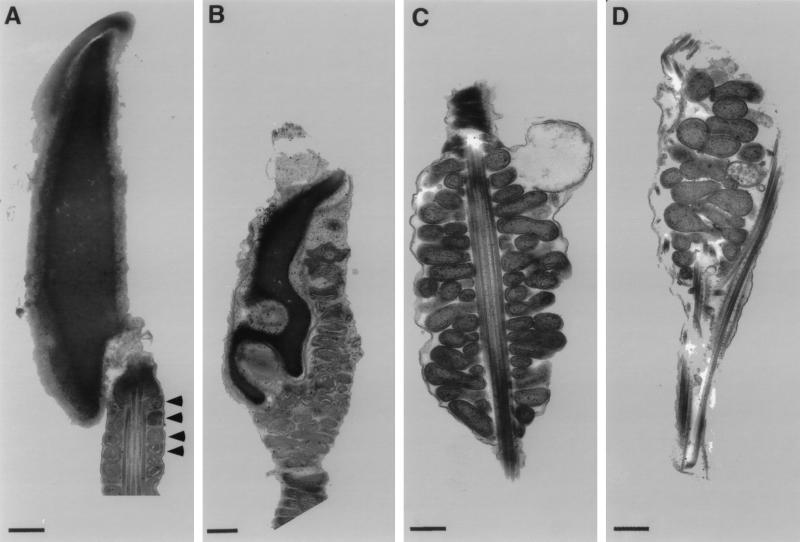
FIG. 3.
Ultrastructure of spermatozoa from wild-type and nectin-2−/− mice. Electron micrographs of spermatozoa from the epididymides and vasa deferentia of nectin-2+/+ (A) and nectin-2−/− (B to D) mice. (A) Head and middle piece. Arrowheads indicate the mitochondrial helical sheath. (B) Head containing mitochondria and deformed nucleus. (C) Deformed mitochondrial helical sheath. (D) Head containing mitochondria and abnormal outer dense fibers. Bars = 500 nm.

