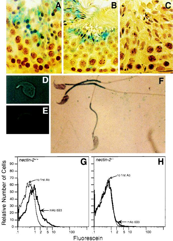
FIG. 5.
Expression of Nectin-2 in germ cells. (A to C) Staining of seminiferous tubules from nectin-2+/+ (A and B) and nectin-2−/− (C) mice with affinity-purified rabbit anti-Nectin-2 antibody. Bound antibody was detected by incubation with biotinylated anti-rabbit antibody followed by avidin–β-galactosidase (41). (A) Stage IX. Staining is predominantly in step 9 spermatids. (B) Stages V and VI. Staining is predominantly in step 15 spermatids. (C) Stages X and XI. No staining is observed in the nectin-2−/− control cells. (D) Light micrograph of isolated condensed spermatid. (E) The same isolated condensed spermatid as in panel D, stained with anti-Nectin-2 monoclonal antibody 6B3 (1) and detected by immunofluorescence microscopy. (F) Spermatozoa from wild-type mice stained with anti-Nectin-2 monoclonal antibody 6B3 and detected by β-galactosidase activity. (G and H) Flow-cytometric analysis of Nectin-2 protein expression on epididymal sperm derived from nectin-2+/+ mice (G) and nectin-2−/− mice (H). Analyses were performed with and without monoclonal antibody (mAb) 6B3. Ab, antibody. Magnifications: ×720 (A to C), ×4,000 (D and E), and ×3,000 (F).

