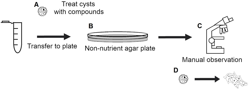
Figure 3. Traditional cysticidal screening workflow.

(A) Cysts are treated and incubated with compounds of interest. (B) Treated cysts are transferred to non-nutrient agar plates with E. coli. (C) Plates are manually imaged and observed daily for evidence of excystation. (D) Observe excystation and proliferation of trophozoites or trails left behind in agar media to manually determine if compound of interest was cysticidal.
