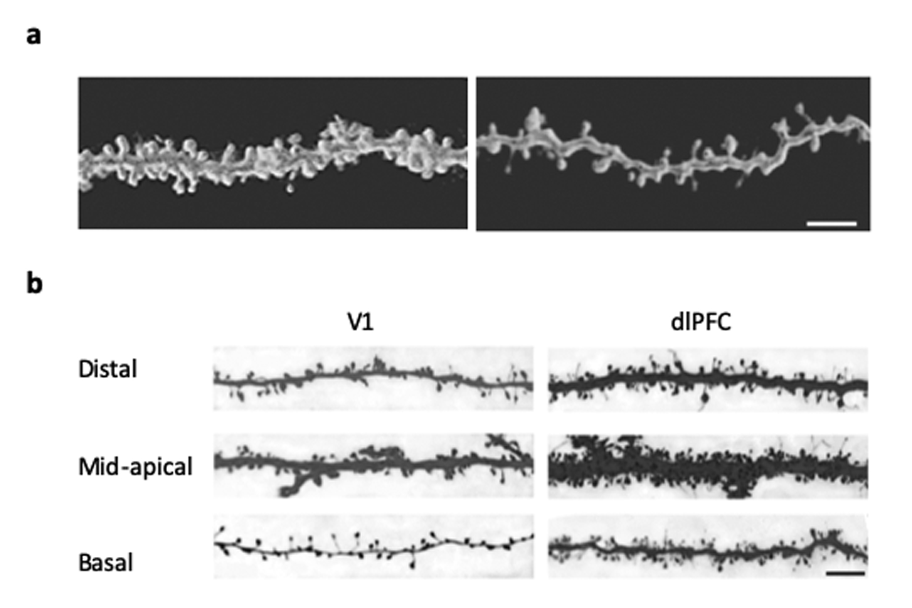
Figure 2. Dendritic spine density and morphology in rhesus macaques.

(A) Confocal image stacks from young (8 years old) (left) vs old (24 years old) (right) macaques show the spine loss associated with aging in dorsolateral prefrontal cortex (dlPFC). Scale bar = 5 μm. (Luebke et al., 2010). (B) Confocal image stacks of distal apical, mid-apical and basal dendritic branches (b/w inverted) showing the fewer dendritic spines and lower proportion of thin spines in visual cortex (V1) when compared to dorsolateral prefrontal cortex (dlPFC) neurons. Scale bar = 5 μm. (Amatrudo et al., 2012)
