


Fig. 2.
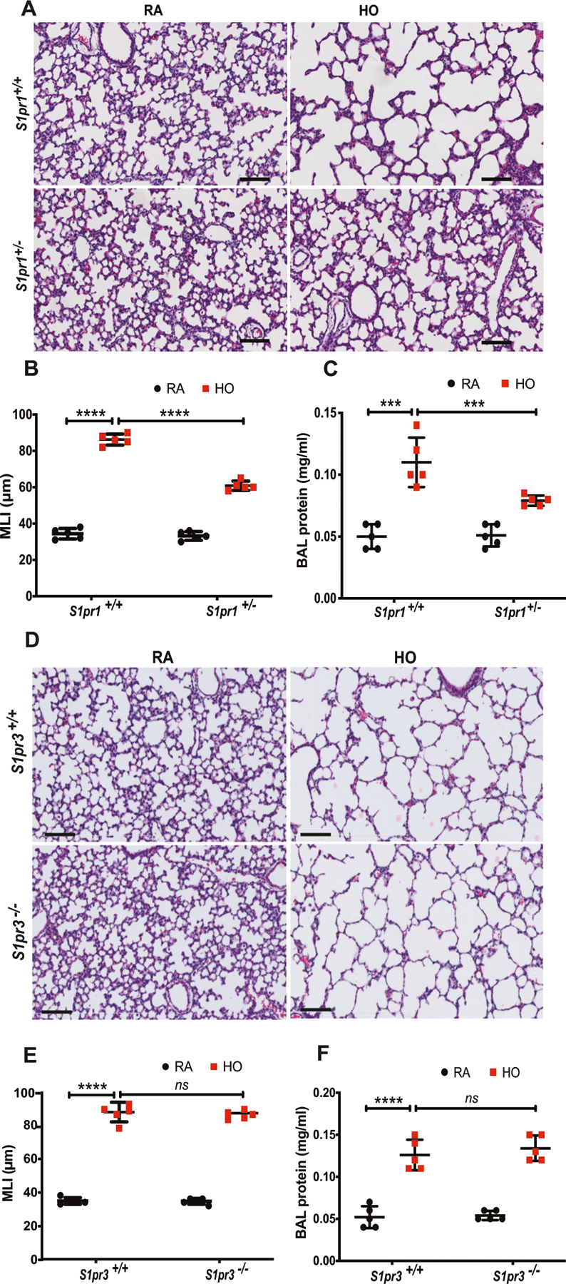
S1pr1 heterozygous (S1pr1+/−) newborn mice showed protection against hyperoxia (HO) induced lung injury whereas S1pr3−/− did not. Representative H&E photomicrographs of lung sections obtained from WT or S1pr1+/− neonatal mice exposed to HO (75% O2) from postnatal day 1 (PN1) for 7 days showed improved alveolarization as compared to S1pr1+/+ controls (A). Objective assessment of alveolarization demonstrated smaller alveoli with shorter mean linear intercept (MLI) in the S1pr1+/− group as compared to controls (B) and less inflammatory exudate in bronchoalveolar lavage fluid (C). Representative H&E photomicrographs of lung sections obtained from WT (S1pr3+/+) or S1pr3−/− neonatal mice exposed to HO (75% O2) from PN1 for 7 days showed no protection against HO-induced BPD as compared to WT controls (D). Objective assessment of alveolarization demonstrated larger alveoli with longer mean linear intercept (MLI) in both the HO-exposed group as compared to RA controls (E) and increased inflammatory exudate in bronchoalveolar lavage fluid (F). Statistical analyses were done with ANOVA. ***p < 0.001, n = 5–8/group