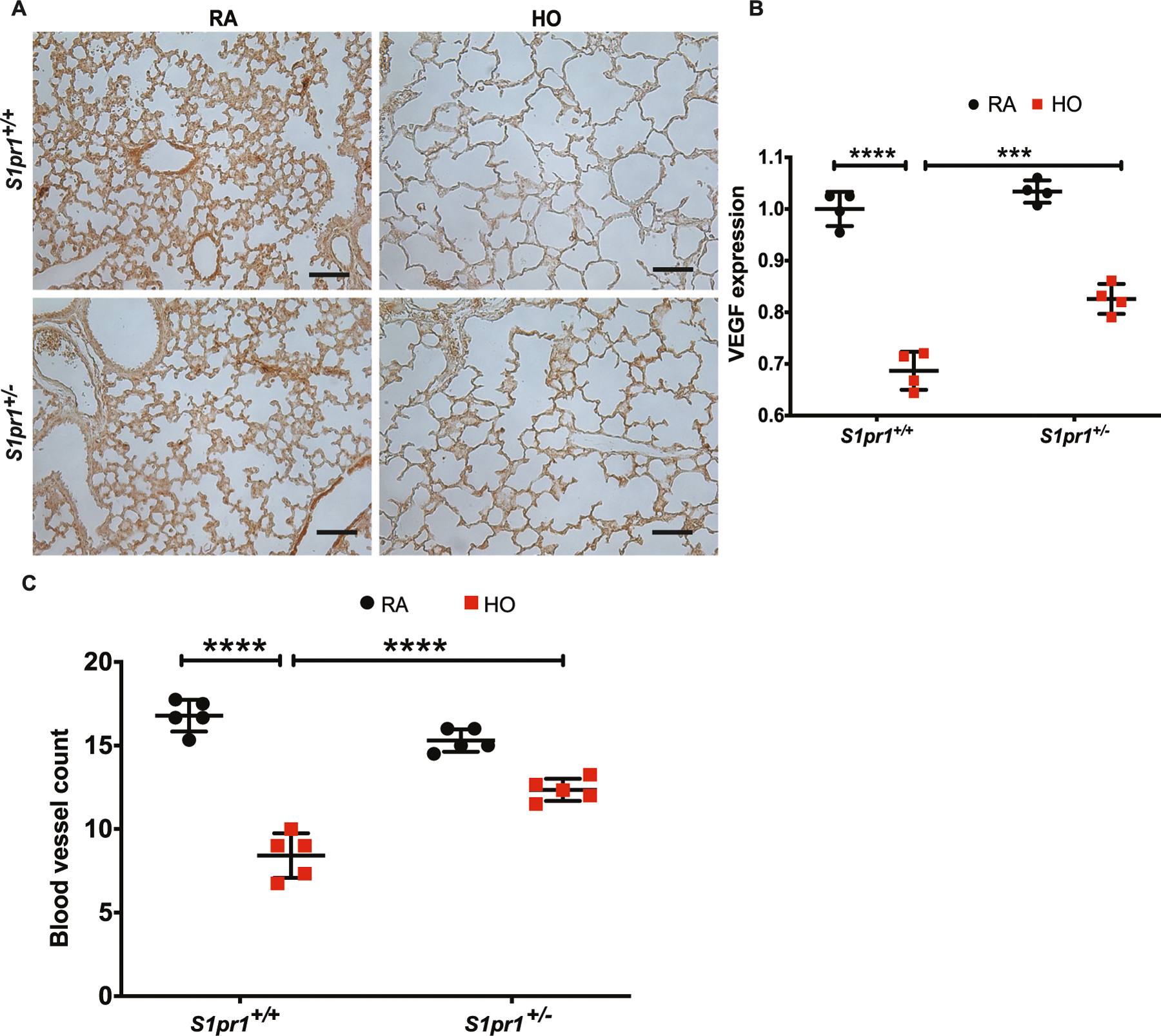
Fig. 4.

Hyperoxia (HO) exposure reduced the expression of VEGF and blood vessel count. HO exposure resulted in reduced VEGF in lung tissue of S1pr1+/+ mice as shown by immunohistochemistry (A, B). This reduction was ameliorated in S1pr1+/− mice which showed higher expression of VEGF. HO was accompanied by a significant reduction in the number of order-1 arterioles in the WT mice which was improved in the S1pr+/− mice (C). The statistical analysis was carried out using ANOVA where *** indicate p < 0.001, n = 5–8/group
