
Figure 3. Dysfunctional SR-BI-mediated DiI-HDL uptake in adipocytes lacking Pcpe2.
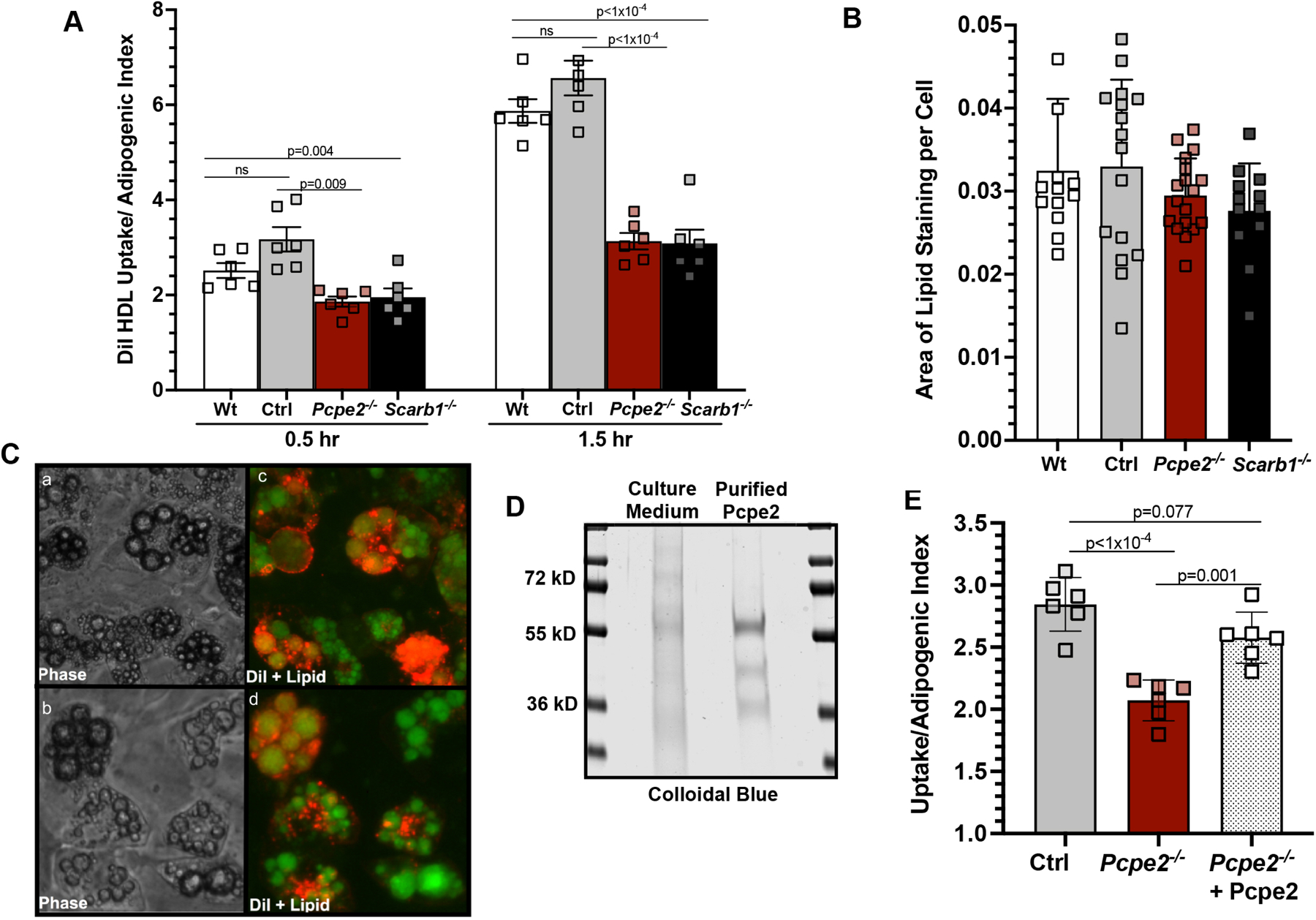
(A) DiI-HDL uptake (10 µg/ml) was measured in day 6 differentiated SVF adipocytes from wild-type (C57BL/6J), Ldlr−/− (Ctrl), Ldlr−/−/Pcpe2−/− (Pcpe2−/−) and Ldlr−/−/Scarb1−/− (Scarb1−/−) cells for 0.5 and 1.5 hours. Uptake normalized to adipogenic index (total area of neutral lipid-stain divided by total DAPI stained nuclei per well). Six wells with 20,000 cells per well were used for each genotype. Data represents average of 4 individual experiments (mean ± SD). (B) adipogenic index for day 6-differentiated SVF adipocytes Wt, Ctrl, Pcpe2−/− and Scarb1−/− mice. (C) Representative (10X) phase contrast (panels a,b) and fluorescent images (panels c,d) of DiI-HDL (red) and bodipy-stained lipid droplets (green) in control (panels a,c) and Pcpe2−/− (panels b,d) cells from day 6 differentiated adipocytes following 1.5 hour incubation with DiI-HDL. (D) SDS- PAGE gel stained with colloidal blue indicates purity of Pcpe2-His protein from 72-hour post-transfection culture medium after Ni-Sepharose purification. Additional details in Supplemental Figure II. (E) Pre-incubation of day 6 differentiated SVF cells lacking Pcpe2 with purified Pcpe2-His protein rescued uptake. Data were analyzed by ANOVA with Tukey’s multiple correction test. lot experiments.