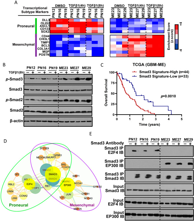
Fig. 2. Smad3 constitutes an important effector in both proneural and mesenchymal GSCs.
A Heatmap showing the molecular subtype marker expression in proneural GSCs (PN12, PN16, and PN19), and mesenchymal GSCs (ME23, ME27, and ME29) with or without TGF-β1 treatment for 4 or 8 h. Proneural markers: DLL3, OLIG2, ASCL1, CD133, and SOX2. Mesenchymal markers: CD44, CHI3L1, TIMP1, BCL3, COL3A1, MGP, and POSTN. Z-scores were calculated from the △Ct values obtained in the quantitative polymerase chain reaction analysis. B Western blotting analysis of SMAD2, p-SMAD2, SMAD3, and p-SMAD3 expression in proneural GSCs (PN12, PN16, and PN19) and mesenchymal GSCs (ME23, ME27, and ME29) with or without TGF-β1 treatment for 8 h. C Kaplan–Meier survival curves for patients with mesenchymal GBM and a high Smad3 gene signature (n = 44) and those with a low Smad3 gene signature (n = 33). D Protein–protein interaction networks indicated by STRING and Cytoscape software show overlapping peaks in both proneural and mesenchymal GSCs and also identify those that are unique to a particular subtype of GSCs or shared between the proneural and mesenchymal GSCs. E Lysates from proneural GSCs and mesenchymal GSCs were subjected to immunoprecipitation using anti-Smad3 antibody and then immunoblotted with anti-E2F4 and anti-EP300 antibodies. GSCs glioblastoma stem cells.

