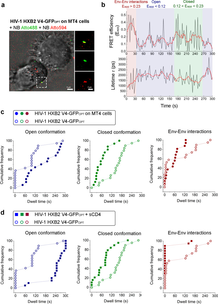
Fig. 3. HIV-1 Env cluster is destabilized when engaged in the pre-fusion reaction in live T cells.
a Micrograph showing mature HIV-1 HXB2 V4-GFPOPT virions labelled with NbA488 (green) NbA594 (red) engaged in the pre-fusion reaction onto living MT4 T cells (phase contrast). The scale bar image on the left is 5 µm. Magnification of the region contoured with dashed lines is shown on the right. Viral particle showing colocalization between green and red channels is shown on the upper right panel. The middle and bottom-right panels correspond to the same viral particle as observed in green and red channels, respectively. Scale bar magnification is 2 µm. b Graphs represent FRET efficiency (Eapp) and lifetime (in ps) traces over time. High FRET efficiency burst (Eapp>0.23), defining intermolecular interactions is depicted in red; intermediate FRET efficiency regime (0.12<Eapp<0.23) assigned to closed Env conformations is depicted in green, and low FRET efficiency bursts (Eapp<0.12) reporting open Env conformations is depicted in blue. Note that high FRET efficiency correlates with low lifetime values and vice versa. c Cumulative Distribution Functions (CDF) are plotted for Eapp single traces obtained from at least (n = 20) HIV-1 HXB2 V4-GFPOPT virions in vitro (open dots) and in presence of living T cells (solid dots). Each FRET regime determines the Env conformational state/Env distribution and kinetics. d Analysis as in (c) of HIV-1 HXB2 V4-GFPOPT virions in vitro (open dots) and in presence of sCD4 (solid squares).

