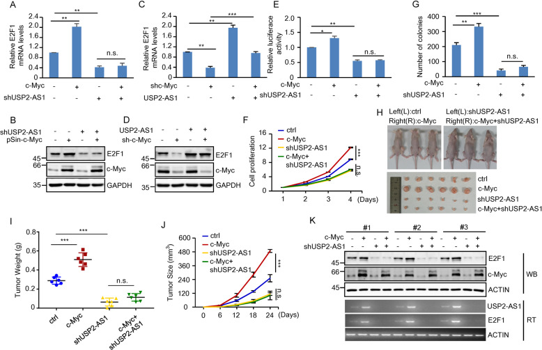
Fig. 7. USP2-AS1 is a mediator of the oncogenic function of c-Myc.
A, B Total RNA (A) and lysates (B) from HCT116 cells expressing control, c-Myc, USP2-AS1 shRNA, or both c-Myc and USP2-AS1 shRNA were analyzed by real-time RT-PCR and western blotting, respectively. **P < 0.01; n.s., no significance. The relative expression levels of USP2-AS1 were also shown in Supplementary Fig. S8A. C, D Total RNA (C) and lysates (D) from HCT116 cells expressing control, c-Myc shRNA, USP2-AS1, or both c-Myc shRNA and USP2-AS1 were analyzed by real-time RT-PCR and western blotting, respectively. **P < 0.01 and ***p < 0.001. The relative expression levels of USP2-AS1 were also shown in Supplementary Fig. S8B. E HCT116 cells expressing control, c-Myc, USP2-AS1 shRNA, or both c-Myc and USP2-AS1 shRNA were transfected with psi-E2F1-3′-UTR. Twenty-four hours later, reporter activity was measured and plotted after normalizing with respect to Renilla luciferase activity. Data shown are mean ± SD (n = 3). *P < 0.05; **p < 0.01; n.s., no significance. F Shown are the growth curves of HCT116 cells expressing control, c-Myc, USP2-AS1 shRNA, or both c-Myc and USP2-AS1 shRNA. Data shown are mean ± SD (n = 3). ***P < 0.001; n.s., no significance. G Colonies of HCT116 cells expressing control, c-Myc, USP2-AS1 shRNA, or both c-Myc and USP2-AS1 shRNA were stained with crystal violet after 8 days of incubation. Data shown are mean ± SD (n = 3). **P < 0.01; ***p < 0.001; n.s., no significance. H–K A total of 2 × 10⁶ HCT116 cells expressing control, c-Myc, USP2-AS1 shRNA, or both c-Myc and USP2-AS1 shRNA were individually injected to the left and right flanks of nude mice as indicated (n = 6 for each group). Representative photographs of mice and xenograft tumors were taken 24 days after injection (H). The excised tumors were weighed (I). Tumor sizes were measured at the indicated time points (J). RNA and protein extracts from the excised xenografts were also analyzed by RT-PCR and western blotting, respectively (K). ***P < 0.001; n.s., no significance. RNA extracts were also analyzed by real-time RT-PCR (Supplementary Fig. S8D).

