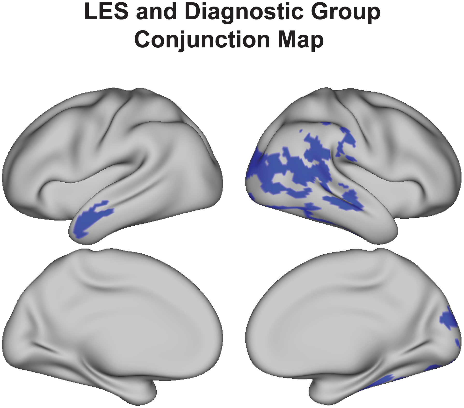
Figure 2.

LES and diagnostic group are both associated with thinner cortex in several of the same cortical areas, including right lateral occipital and temporal cortex and the left lateral anterior temporal lobe. Conjunction map indicates vertices (shown in blue) which are sensitive to both LES (Figure 1a) and diagnostic group (Figure 1b), separately.
