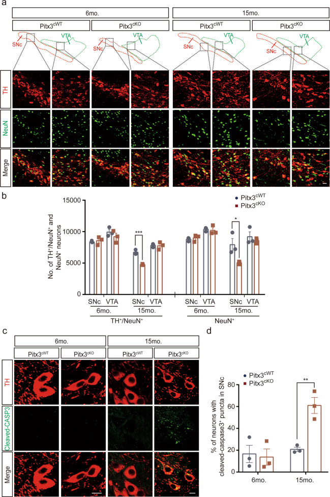
Fig. 3. Neurodegeneration in 15-month-old Pitx3cKO mice accompanied by the promoted apoptosis.
a IFC co-staining of TH and NeuN, and IFC staining of NeuN in the ventral midbrain sections from 6 and 15-month-old Pitx3cWT and Pitx3cKO mice. SNc and VTA were outlined, respectively (scale bar: 10 μm). b Quantification of TH+/NeuN+ and NeuN+ neurons in the SNc and VTA from 6- and 15-months-old Pitx3cWT and Pitx3cKO mice (N = 3 mice per genotype). unpaired t-test, ***p = 0.0008 (15 months old for TH+/NeuN+ co-staining); unpaired t-test, *p = 0.0424 (15 months old for NeuN+ staining). c IFC staining of cleaved-caspase3 in the SNc area from 6 and 15-month-old Pitx3cWT and Pitx3cKO mice (scale bar: 10 μm). d Quantification of the percentage of neurons with cleaved-caspase3+ puncta in SNc (N = 3 mice per genotype). unpaired t-test, **p = 0.0062.

