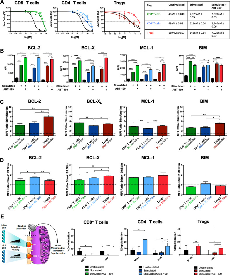
Fig. 1. Ex vivo stimulation and expansion of CD8+ T cells, CD4+ T cells, and Tregs alone or in the setting of BCL-2 blockade results in divergent cell death resistance and BCL-2 family protein expression.
A Cell death of CD8+ T cells, CD4+ T cells, and Tregs treated with venetoclax directly following isolation and after stimulation/expansion alone or in the presence of venetoclax. B BCL-2 family protein levels of unstimulated and stimulated/expanded T cells alone or in the presence of venetoclax. C Ratios of BCL-2 family protein levels in stimulated/expanded T cells compared to unstimulated and D between stimulated/expanded in the presence of venetoclax compared to stimulated/expanded alone. E BH3 profiling of T cells using peptides specific for MCL-1 (NOXA) and BCL-XL (HRK) to determine shifts in anti-apoptotic dependencies. Schematic of BH3-peptide specificity and ability to induce mitochondrial outer membrane permeabilization (MOMP) is shown on the left-hand side. Data represented in black represents treatment of cells prior to stimulation. Colored data (CD8+ T cells in green, CD4+ T cells in blue, and Tregs in red) represent treatment following stimulation (dark green, blue, and red) or following stimulation in the presence of venetoclax (light green, blue, and red). Data is representative of three independent replicates. Data represented as means ± SEM. *p < 0.05, **p < 0.01, ***p < 0.001, ****p < 0.0001.

