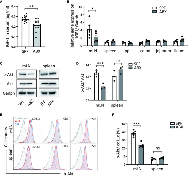
Figure 2.
Gut dysbiosis regulates the IGF-1/Akt pathway in mesenteric lymph tissue. (A) After mice treated with ABX, IGF-1 ELISA was performed on serum. (B) And mRNA expression of IGF-1 in mLN, spleen, Peyer’s Patches (PP), colon, jejunum, ileum tissues, were measured by RTPCR. (C, D) p-Akt and Akt protein level change in mLN and spleen were determined by western blotting. (E) p-Akt expression levels of DCs (cd11c+), T cells (CD3+), B cells (B220+) in mLN and spleen were measured by flow cytometry. (F) Graph shows frequency of p-Akt positive DCs in mLN and spleen. As shown, p-Akt protein level of DC in mLN was reduced in ABX mice. Graph shows mean and SEM. (A) Data are combined from two independent experiments with at least thirteen mice per group. Graph shows mean and SEM. Data were analyzed by unpaired Student’s t-test. *p < 0.05, **p < 0.01, ***p < 0.001, ns, not significant.

