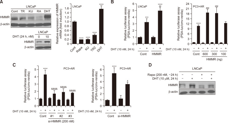
Fig. 5.
Androgen induces HMMR expression by activating mTOR, and silencing HMMR suppresses androgenic responses in LNCaP cells. (A) LNCaP cells were cultured for 24 h with the indicated pharmacological reagents before lysis (TR; Torin2, KU; KU0063794, RA; Rapamycin, DHT; Dihydrotestosterone). Protein samples and total RNA isolated were subjected to immunoblotting and quantitative real time PCR, respectively to measure HMMR expression. Data shown are the mean ± SD of triplicate determinations and representative of two or three independent experiments (****p<0.0001 vs.Cont). (B) Cells were transiently transfected with 1 μg of total DNA including PSA-luciferase (PSA-luc) construct, cmv-renilla and either pCMV6 or pCMV6-flag-HMMR, followed by DHT (10 nM) treatment for 24 h (***p<0.001, ****p<0.0001 vs. Cont, ##p<0.01, ###p<0.001 vs. DHT alone). (C) Separately, cells were transiently transfected with 1 μg of total DNA including either PSA-luciferase (PSA-luc) or MMTV-luc construct and cmv-renilla, and 200 nM of si-Cont or si-HMMR. Cells were then cultured for 24 h in the absence or presence of DHT (10 nM). Data shown are relative values of firefly luciferase activity normalized to renilla luciferase. Data shown are the mean ± SD of triplicate determinations and representative of two or three independent experiments (****p<0.0001 vs.Cont, #p<0.05, &&&&p<0.0001 vs. DHT alone). (D) LNCaP was stimulated with 200 nM Rapamycin for 24 h, followed by DHT treatment for additional 24 h. The expression level of HMMR protein was measured by western blot analysis. Data shown are the mean ± SD of triplicate determinations and representative of two or three independent experiments (B-D).

