
Figure 2.


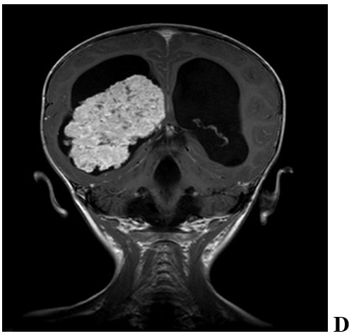


Radiographic features of a pediatric CPP in 2 year old female. We found significant hydrocephalus (A-F) with transependymal flow (F) due to either increased production of CSF or obstruction by tumor debris and blood products. Intraventricular and/or intratumoral hemorrhage are not uncommon. CPPs enhance on T1-weighted MRI with contrast (C,D and E) due to their rich vascular supply. They are generally iso-or hypointense on T1-and T2-weighted MRI (A, B and F) but may demonstrate heterogeneous hyperintensity in some cases. Magnetic resonance angiography may aid in visualizing the tumor’s vascular supply (C, D and F)