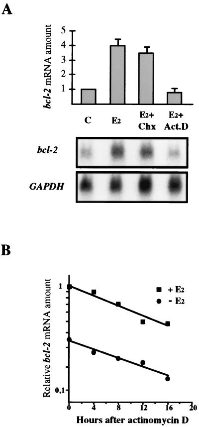
FIG. 1.
Effect of cycloheximide and actinomycin D on bcl-2 mRNA up-regulation by 17β-estradiol. (A) On top are graphically represented the results of Northern blot analysis carried out with MCF-7 cells depleted by the hormone for 7 days (C) or treated for 18 h with 10 nM 17β-estradiol alone (E2) or with 25 μg of cycloheximide per ml (E2+Chx) or 1 μg of actinomycin D per ml (E2+Act.D). Amounts of bcl-2 mRNA are reported relative to that measured in starved cells (C), set to 1. Twenty micrograms of total RNA was loaded onto each lane and run on a 1% agarose gel before blotting and hybridization. bcl-2 mRNA was quantified by phosphorimaging analysis and normalized to GAPDH. The error bars represent the standard errors of two different experiments. The observed fourfold increase of bcl-2 mRNA induced by the hormone was inhibited only by actinomycin D treatment. At the bottom is reported the result of one experiment after a 15-day exposure. (B) Hormone-depleted MCF-7 cells were incubated with or without 10 nM 17β-estradiol for 24 h; actinomycin D (2 μg/ml) was then added for the reported times prior to RNA extraction. Twenty micrograms of total RNA per sample was then processed as described above. RNA amounts are represented as fractions of that assayed at time zero in hormone-challenged MCF-7 cells, which was given the arbitrary value of 1. Treatment with 17β-estradiol did not affect bcl-2 mRNA stability since, after 16 h of actinomycin D addition, roughly 40% of the initial RNA amount was measured in both hormone-challenged and -depleted cells.

