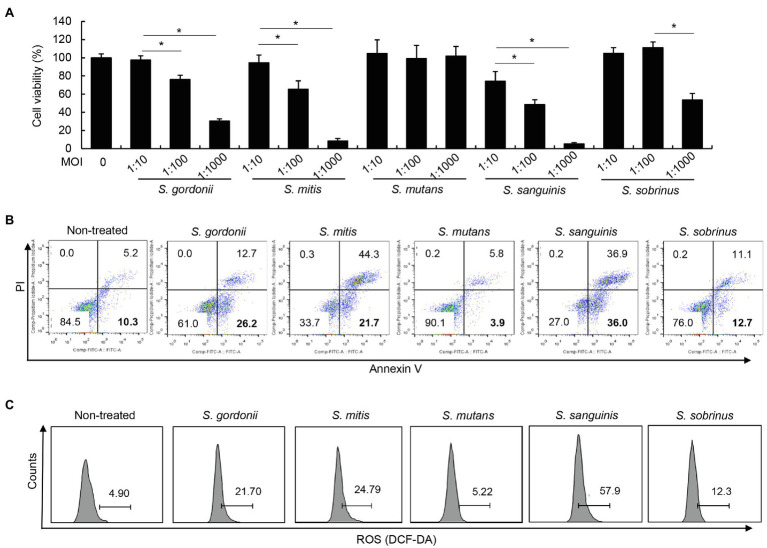
Figure 4.
Various streptococcal species differ in induction of apoptosis and ROS in human PDL cells. (A) PDL cells were treated with S. gordonii, S. mitis, S. mutans, S. sanguinis, and S. sobrinus at MOI 1:10, 1:100, or 1:1,000 for 3h. Trypan blue assay was used to determine the number of viable cells. (B) PDL cells were treated with S. gordonii, S. mitis, S. mutans, S. sanguinis, and S. sobrinus at MOI 1:1,000 for 1h. The cells were strained with annexin V and PI and then analyzed using flow cytometry. (C) PDL cells were treated with 10μM of DCF-DA for 30min at 37°C. The DCF-DA-treated cells were washed with PBS and then treated with S. gordonii, S. mitis, S. mutans, S. sanguinis, and S. sobrinus at MOI 1:1,000 for 3h in a CO2 incubator. Fluorescent intensity was analyzed by flow cytometry. One of three similar results is shown. *p<0.05.

