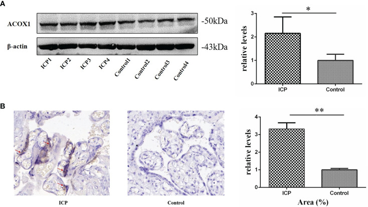
Figure 2.
Validation of ACOX1 protein in placental tissue. (A) Western blot analysis of placental ACOX1 in tissue obtained from pregnant women with ICP and healthy pregnant women. Placental ACOX1 levels were significantly increased among pregnant women with ICP as compared to healthy pregnant women (P=0.015); β-actin was used as an internal control. (*P < 0.05); (B) Immunohistochemical staining for placental ACOX1 among 4 pregnant women with ICP and 4 healthy pregnant women (×400). Immunohistochemistry revealed higher cytoplasmic and nuclear levels of ACOX1 in placental trophoblasts of pregnant women with ICP as compared to healthy pregnant women. The mean positive signal intensity of placenta in ICP group was significantly higher than that in control group (**P < 0.01).

