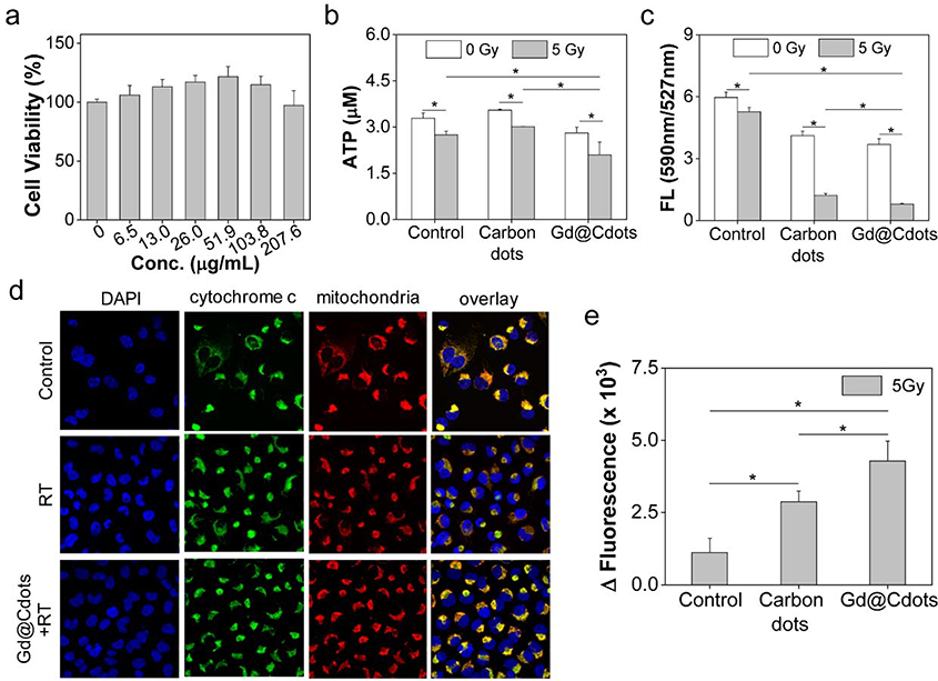
Figure 5.
Impact of Gd@Cdots on cell viability, evaluated with H1299 cells. (a) Cell viability in the absence of radiation, measured by MTT assay. (b) Cell viability under radiation (5 Gy), measured by ATP bioluminescence assay. Gd@Cdots (30 μg/mL) were incubated with cells during radiation. Cdots were also tested as a comparison. (c) Mitochondrial membrane potential change, measured by JC-1 staining. (d) Cytochrome c release, evaluated by cytochrome c and mitochondria double staining. Cytochrome c translocation into the cytosol was indicated by red arrows. (e) Activation of apoptosis, evaluated by caspase 3 activity assay. *, p < 0.05.

