
Fig. 2-.
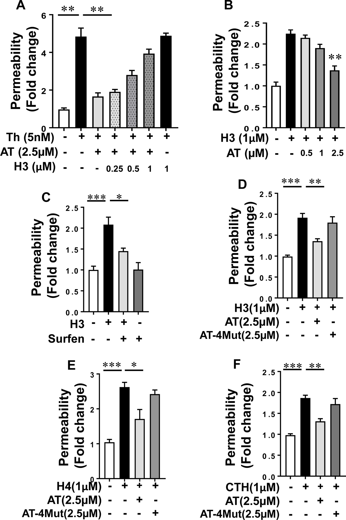
Histone H3 inhibits the endothelial barrier protective function of AT. (A) Confluent HTERT-HUVECs were simultaneously incubated with a fixed concentration of AT (2.5 µM) and increasing concentration of histone H3 for 4h followed by measuring cell permeability in response to thrombin by spectrophotometric measurement of the flux of Evans blue-bound albumin across functional endothelial cell monolayer as described in methods. (B) The same as (A) except that the fold change in cell permeability was measured by simultaneous incubation of a fixed concentration of histone H3 with increasing concentration of AT. (C) The cell permeability induced by histone H3 (1 µM for 4h) was monitored in the absence and presence of the GAG-antagonist surfen (10 µM). (D) Protective effect of WT-AT and its D-helix mutant (AT-4Mut) on histone H3-mediated endothelial cell permeability was measured by influx of albumin-bound Evans blue across functional endothelial cell monolayer. (E) The same as (D) except that the effect of WT-AT and AT-4Mut on histone H4-mediated permeability was monitored. (F) The same as (D) except that the effect of WT-AT and AT-4Mut on calf thymus histone (CTH)-mediated cell permeability was monitored. Neither AT-WT nor AT-4Mut had an effect on cell permeability in the absence of histones (not shown). All results are shown as means ± SD of three different experiments. * p < 0.05, ** p < 0.01, *** p < 0.001.