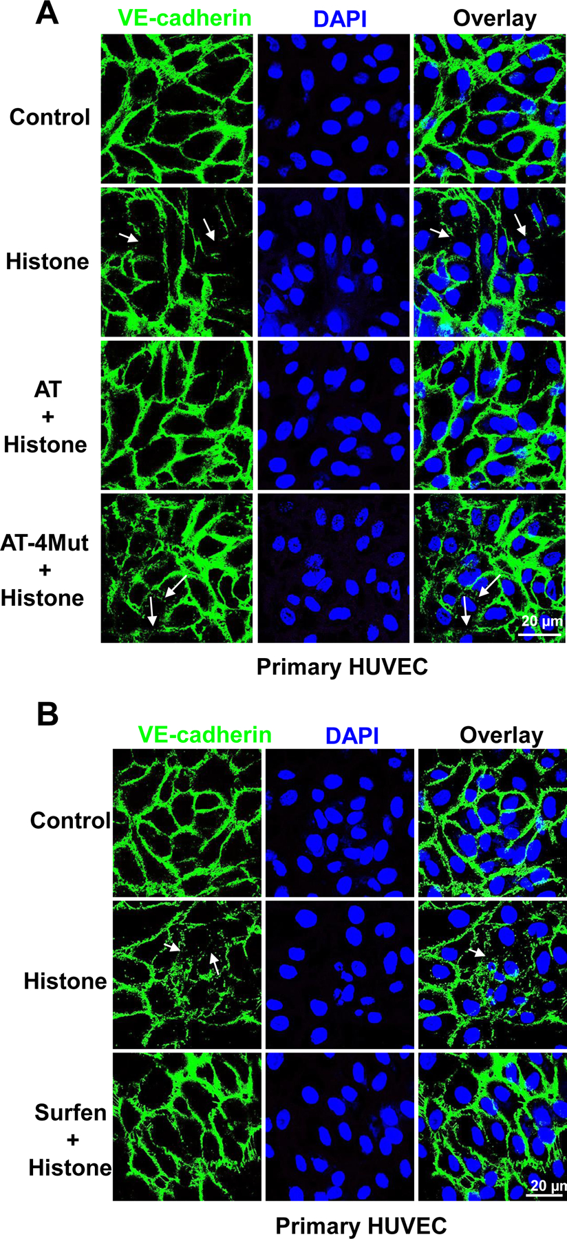
Fig. 4-.

Immunofluorescence analysis of histone-mediated VE-cadherin disruption in endothelial cells. (A) Primary HUVECs were incubated with Histone H3 (1 µM) for 1h in the absence or presence of WT-AT and AT-4Mut (2.5 µM each). Cells were then fixed, permeabilized and incubated with rabbit anti-VE-cadherin antibody and Alexa Fluor 488-conjugated goat anti-rabbit IgG. The nucleus was stained with DAPI. Immunofluorescence images were obtained with confocal microscopy. (B) The same as (A) except that primary HUVECs were incubated with histone H3 for 1h in the absence or presence of surfen (10 µM). Arrows indicate loss of VE-cadherin at junctions.
