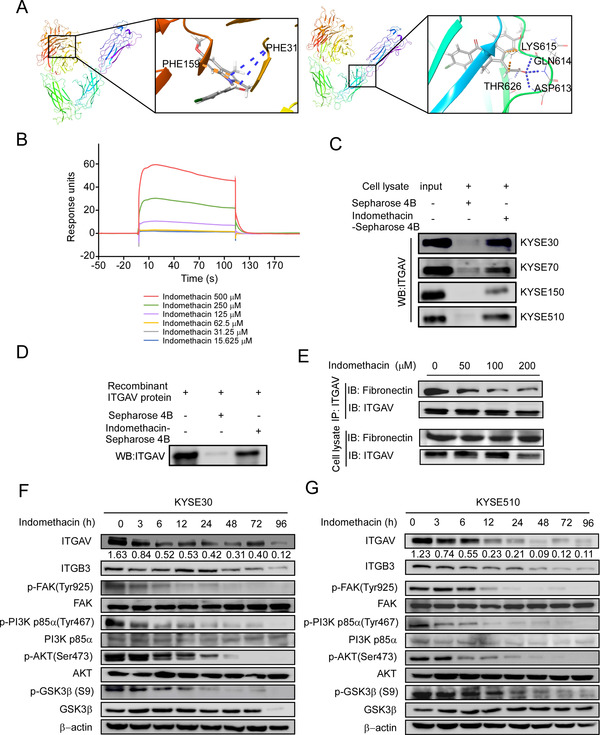
FIGURE 3.

Indomethacin binds to ITGAV. (A) Computational modelling illustrating binding between indomethacin and ITGAV. Indomethacin binds to ITGAV at PHE31 and PHE159 (upper panel) or LYS615, GLN614, and ASP613 (lower panel). The ITGAV structure is visualized as a ribbon; indomethacin is visualized as a stick. (B) Binding sensorgrams for the interaction between indomethacin and immobilized ITGAV. The ITGAV immobilization level was 200 RU. ESCC cell lysates (C) or ITGAV recombinant proteins (D) were mixed with 50 µl of indomethacin‐conjugated Sepharose 4B beads or DMSO‐conjugated Sepharose 4B beads in reaction buffer. The pulled‐down proteins were detected by Western blotting. (E) ITGAV‐overexpressing HEK‐293T cells were lysed and added to 20 µg of fibronectin proteins. After incubation with the indicated concentrations of indomethacin at 4°C overnight, proteins were subjected to IP assay. (F and G) ITGAV, ITGB3, p‐FAK, FAK, p‐PI3K, PI3K, p‐AKT, AKT, p‐GSK3β, and GSK3β protein levels were assessed in KYSE30 and KYSE510 cells by Western blotting after treatment with DMSO or 200 µM indomethacin (0–96 h). For (B–G), three independent experiments were performed
