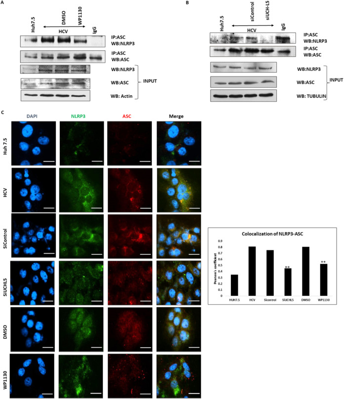
FIG 4.
Effect of UCHL5 in NLRP3 inflammasome formation during HCV infection. (A) Huh7.5 (mock) and HCV-infected cells 7 days postinfection (p.i.) were treated with WP1130 (5 μM) for 4 h and lysed in NP-40 lysis buffer. ASC was immunoprecipitated using anti-ASC antibody and Western blotted for NLRP3 and ASC. Input samples were blotted for NLRP3, ACS, and actin. (B) HCV-infected cells were transfected with control or UCHL5 siRNA at 4 days p.i. using electroporation and were lysed 7 days p.i. in NP-40 lysis buffer. ASC was IP using anti-ASC antibody and Western blotted for NLRP3 and ASC. Input samples were blotted for NLRP3, ASC, and tubulin. Representative images are presented from three independent experiments for panels A and B. (C) Mock, HCV-infected, and HCV-infected samples treated with WP1130 (5 μM) or transfected with siUCHL5 were fixed using 4% paraformaldehyde. The cells were washed with phosphate-buffered saline (PBS) and were permeabilized using 0.2% Triton X-100. The cells were washed and blocked using Image-iT FX signal enhancer for 20 min. The cells were then incubated for 2 h using primary antibody for NLRP3 and ASC. After incubation with primary antibody, the cells were washed again and incubated for 2 h with Alexa Fluor 488 and Alexa Fluor 594 secondary antibody for NLRP3 and ASC, respectively. After washing with PBS, the cells were mounted using mounting medium containing DAPI and imaged using a Nikon i80 microscope (scale bars are 20 μm). Mean Pearson’s coefficients were calculated using ImageJ for three fields for each sample containing approximately 50 cells/field. Error bars are the standard deviation from the mean, and significance (P < 0.05 [**]) was calculated using a student’s t test. Similar results were obtained in two biological replicates.

