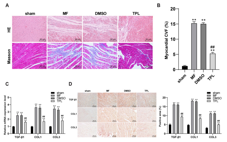
Fig. 3. TPL downregulated the degree of MF and the expression of fibrosis related factors in MF rats.
(A) H&E staining and Masson staining were used to observe the pathological morphology and MF of rats in each group (×200); (B) Masson staining results were quantitatively analyzed by measuring collagen volume fraction; (C) RT-qPCR was used to detect the expressions of TGF-β1, COL1, and COL3; (D) The expressions of TGF-β1, COL1, and COL3 were detected by immunohistochemistry; n = 6. Three independent repeated tests were performed and the data were expressed as mean ± SD; one-way was used for variance analysis; Tukey’s multiple comparisons test was used for post-hoc test. TPL, triptolide; MF, myocardial fibrosis; DMSO, dimethyl sulfoxide. Compared with sham group, **p < 0.01; compared with MF group, #p < 0.05, ##p < 0.01.

