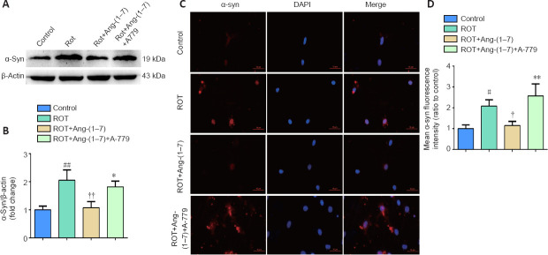
Figure 5.
Ang-(1–7) attenuates α-synuclein accumulation in the rotenone-induced cell model in a MasR-dependent manner.
(A) The expression of α-syn in four groups was examined by Western blot assay. (B) Quantitative results of α-syn expression. (C) Cells were marked by an anti-α-syn antibody (red), nuclei were counterstained with DAPI (blue), and immunofluorescence was observed by fluorescent microscopy (original magnification, 630×). Expression of α-syn was higher in the ROT group and was downregulated when incubated with Ang-(1–7). However, the influence caused by Ang-(1–7) was completely abolished with A-779 co-treatment. (D) Mean α-syn fluorescence intensity (ratio to control) of four groups. Data are expressed as the mean the ± SD (n = 3; one-way analysis of variance followed by Tukey's post hoc test). #P < 0.05, ##P < 0.01, vs. control group; †P < 0.05, ††P < 0.01, vs. ROT group; *P < 0.05, **P < 0.01, vs. ROT + Ang-(1–7) group. Ang-(1–7): Angiotensin-(1–7); DAPI: 4′,6-diamidino-2-phenylindole; MasR: Mas receptor; ROT: rotenone; α-syn: α-synuclein.

