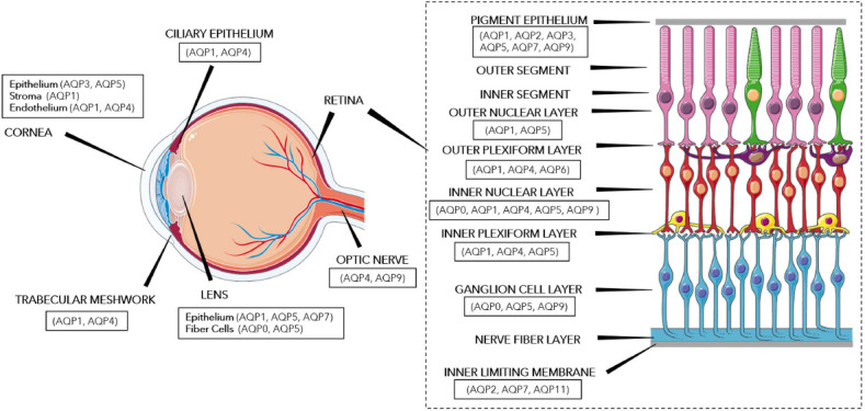
Figure 1.

Aquaporin distribution in ocular tissues.
This diagram portrays the aquaporins expressed in various ocular tissues, including the cornea, ciliary epithelium, trabecular meshwork, lens, optic nerve, and the various layers of the retina. Aquaporin distribution data was adapted from Schey et al. (2014) and images of the eye and the retina were modified from SMART (Servier Medical Art), licensed under a Creative Common Attribution 3.0 Generic License. http://smart.servier.com/. AQP: Aquaporin.
