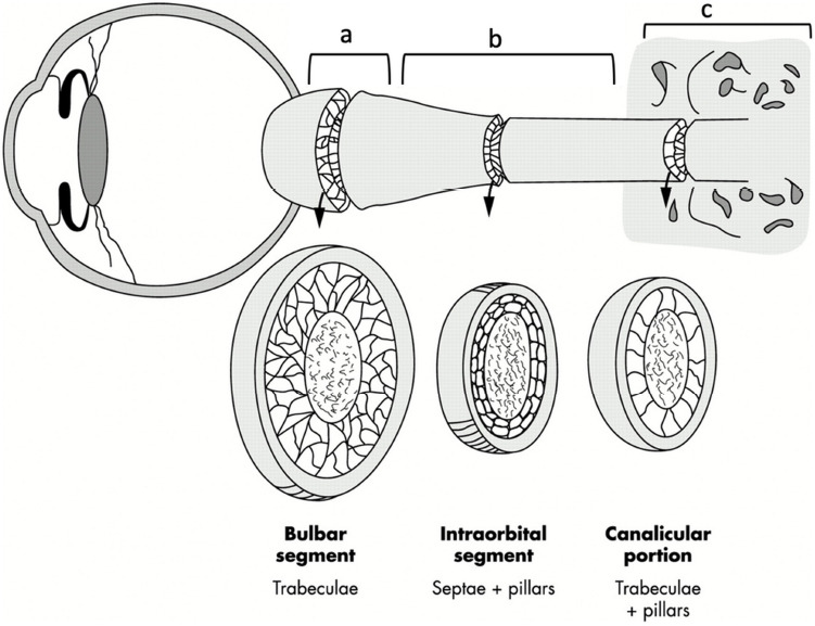
Figure 2.

Anatomy of optic nerve subarachnoid space.
Schematic drawing of the optic nerve demonstrating the location of the (a) bulbar segment, (b) mid-orbital segment, and (c) canalicular portion. The bulbar segment (a) and the mid-orbital segment (b) together form the orbital portion. The SAS in the bulbar segment is composed of trabeculae, the SAS in the mid-orbital segment mainly comprises the septae and pillars, and the SAS in the canalicular portion contains both trabeculae and pillars. Reproduced with permission from Killer et al. (2003). SAS: Subarachnoid space.
