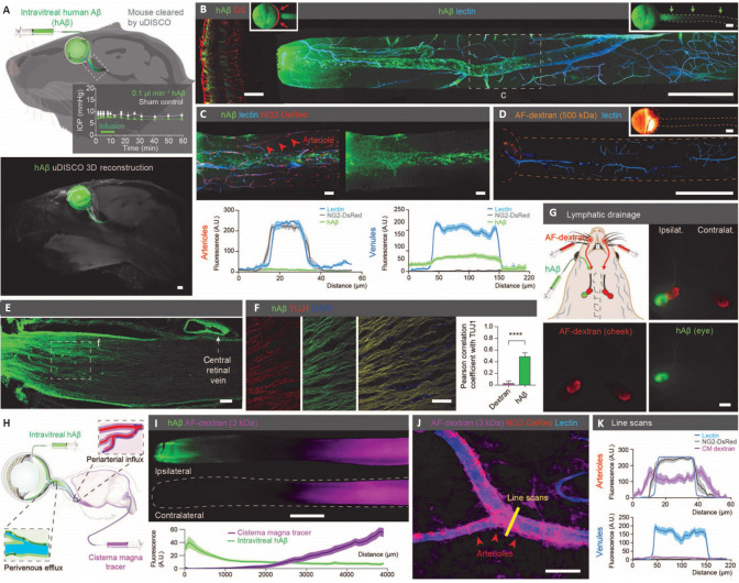
Figure 3.
Ex vivo imaging of the glymphatic system in the optic nerve.
(A) The top image visualizes the intravitreal injection of human amyloid beta (hAß); the graph displays the mouse intraocular pressure (IOP) stabilizing during injection; the bottom image displays the uDISCO-cleared transparent mouse heads 1 hour after hAβ injection, showing that hAβ tracer exited the eye along the optic nerve. (B) These confocal images of ipsilateral retina (left) and optic nerve (right) after intravitreal hAβ injection confirmed that hAβ tracer is transported anterogradely along the nerve. (C) Confocal images from reporter mouse with DsRed-tagged mural cells 30 minutes after intravitreal hAβ injection showed that hAβ tracer preferentially accumulated in the perivascular space along the optic nerve veins. (D) Confocal image of mouse optic nerve 30 minutes after intravitreal Alexa Fluor-dextran injection showed no entrance of this tracer into the nerve. (E, F) Confocal images of optic nerve co-labeling with TUJ1 after tracer administration. Note tracer accumulation in the dural lining of the nerve. (G) The cervical lymph nodes are exhibiting intense hAβ labeling 3 hours after injection. (H) Schematic of double injections of hAβ intravitreally and fluorescent tracer intercisternally. (I) Representative image and quantification of the double injections described in (H), which highlights that tracers transported within the optic nerve in both anterograde and retrograde directions. (J) Confocal images of the optic nerve from reporter mouse with DsRed-tagged mural cells (vascular smooth muscle cells and pericytes) after intracisternal dextran injection with line scan quantified in (K). (J) and (K) show that the tracer injected intercisternally predominantly transported along the periarterial and pericapillary spaces. Reproduced with permission from Wang et al. (2020). AF: Alexa Fluor; DAPI: 4′,6-diamidino-2-phenylindole; DsRed: Discosoma sp. red fluorescent protein; GS: glutamine synthetase; hAß: human amyloid beta; IOP: intraocular pressure; NG2: neuron-glial antigen 2; TUJ1: neuron-specific class III beta-tubulin; uDISCO: ultimate 3D imaging of solvent-cleared organs.

