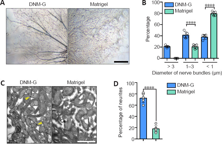
Figure 1.

Morphological analysis of axons in a peripheral nerve ECM-derived environment.
(A) DRG axons growing on different ECM substrates. The DNM-G substrate induced the formation of axon bundles, whereas the Matrigel gave rise to dispersed axons. (B) We compared the distribution of axon bundle diameters between DNM-G and Matrigel treated DRGs (n = 5 cultures, ****P < 0.0001, Student's t-test). (C) Electron microscopy of axons in DRG ex vivo preparations. There were more axonal contacts in the DNM-G group than in the Matrigel group. Arrowheads indicate contacts between axons. (D) Percentage of axons attached to one another in the DNM-G and Matrigel groups (n = 6 independent experiments, ****P < 0.0001, Student's t-test). Scale bars: 600 μm in A and 1 μm in C. DNM-G: Decellularized nerve matrix-gel; DRG: dorsal root ganglion; ECM: extracellular matrix.
