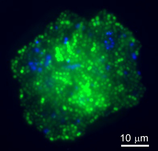
FIG 1.

Visualization of coaggregation between Streptococcus gordonii and Streptococcus oralis. Example of a coaggregate formed between S. gordonii (Syto 9; green) and S. oralis (4′-6-diamidino-2-phenylindole [DAPI]; blue) in 25% human saliva. Cells were prestained before mixing and were visualized by fluorescence microscopy. The image shows a large aggregate.
