Figure 3.
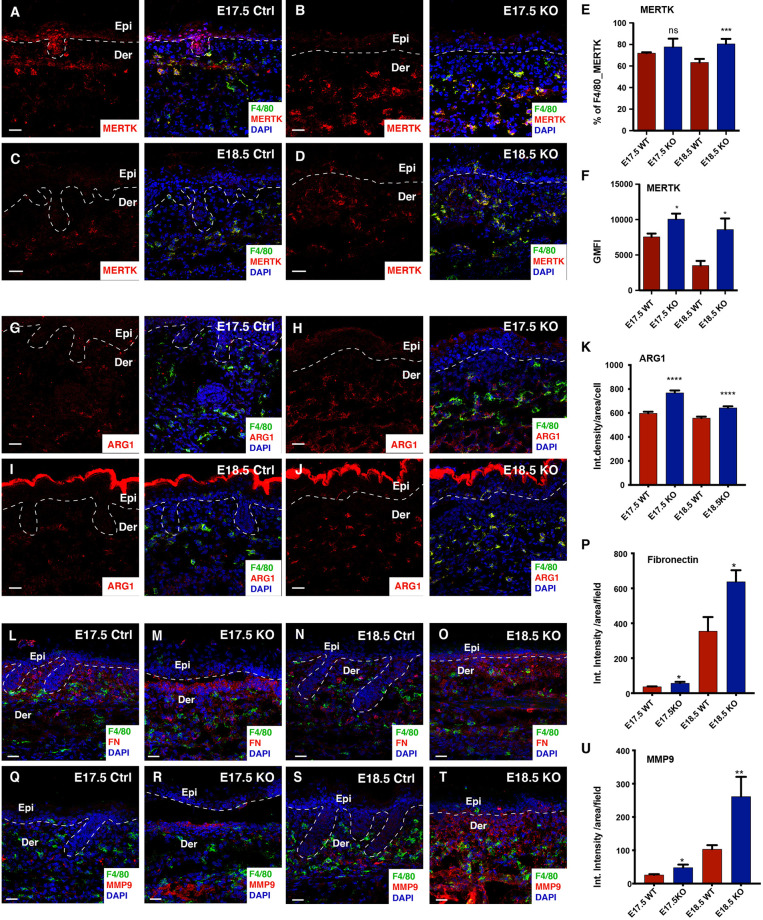
Macrophages in the KO skin acquire exaggerated M2-like pro-remodelling properties at E18.5. Immunostaining in the control and Itgβ1 epidermal KO skin at E17.5 and E18.5 for F4/80 and MERTK (A–D) quantified in (E) (N=3). Surface level expression of MERTK is quantified (F) (N=3). Immunostaining in the control and Itgβ1 epidermal KO skin at E17.5 and E18.5 for F4/80 and Arg1 (G–J) quantified in (K) (N=2). Immunostaining in the control and Itgβ1 epidermal KO skin at E17.5 and E18.5 for F4/80 and FN1 (L–O) quantified in (P) (N=2). Immunostaining in the control and Itgβ1 epidermal KO skin at E17.5 and E18.5 for F4/80 and MMP9 (Q–T) quantified in (U) (N=2). The white dashed line separates the epidermis (Epi) and the dermis (Der). Scale bars: 20 um. (*p ≤ 0.05, **p ≤ 0.01, ***p ≤ 0.001, ****p ≤ 0.0001, ns=not significant).

