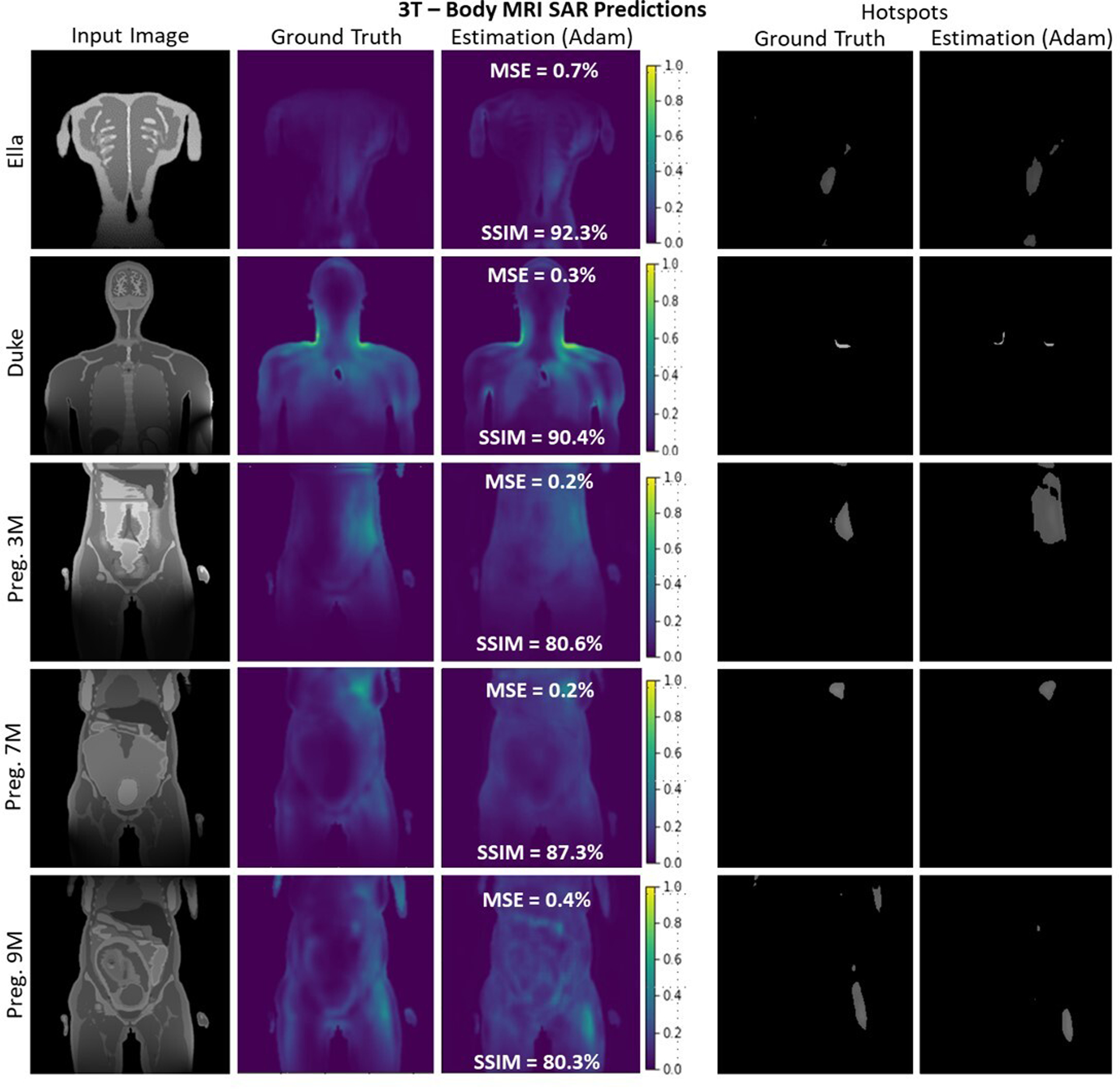
FIGURE 5.

A selection of slices for Ella, Duke, and Pregnant Women of different gestational stages at 3T body imaging. Despite very high variation of SAR maps and input images, the proposed 4-stage U-Net architecture successfully recovered the distribution with better than 80% average SSIM and less than 0.5% average MSE for all body models.
