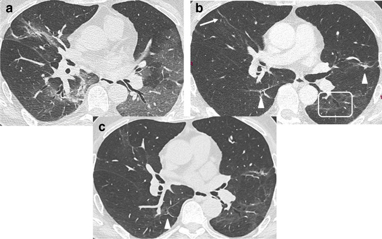
Fig. 4.
CT features at baseline and 4-month and 1-year follow-up, in a 65-year-old man who developed severe COVID-19 pneumonia (intubation, 27 days in ICU). a Baseline CT. b 4-month CT follow-up. Ground glass opacities (GGOs) have largely resolved, except in the upper segment of the left lower lobe. Bronchial dilatation (square) is demonstrated within the residual GGO area. Linear consolidation (arrow head) and parenchymal band (arrow) are also denoted. c 1-year follow-up. CT features are unchanged. There are no honeycombing and no peripheral reticulations

