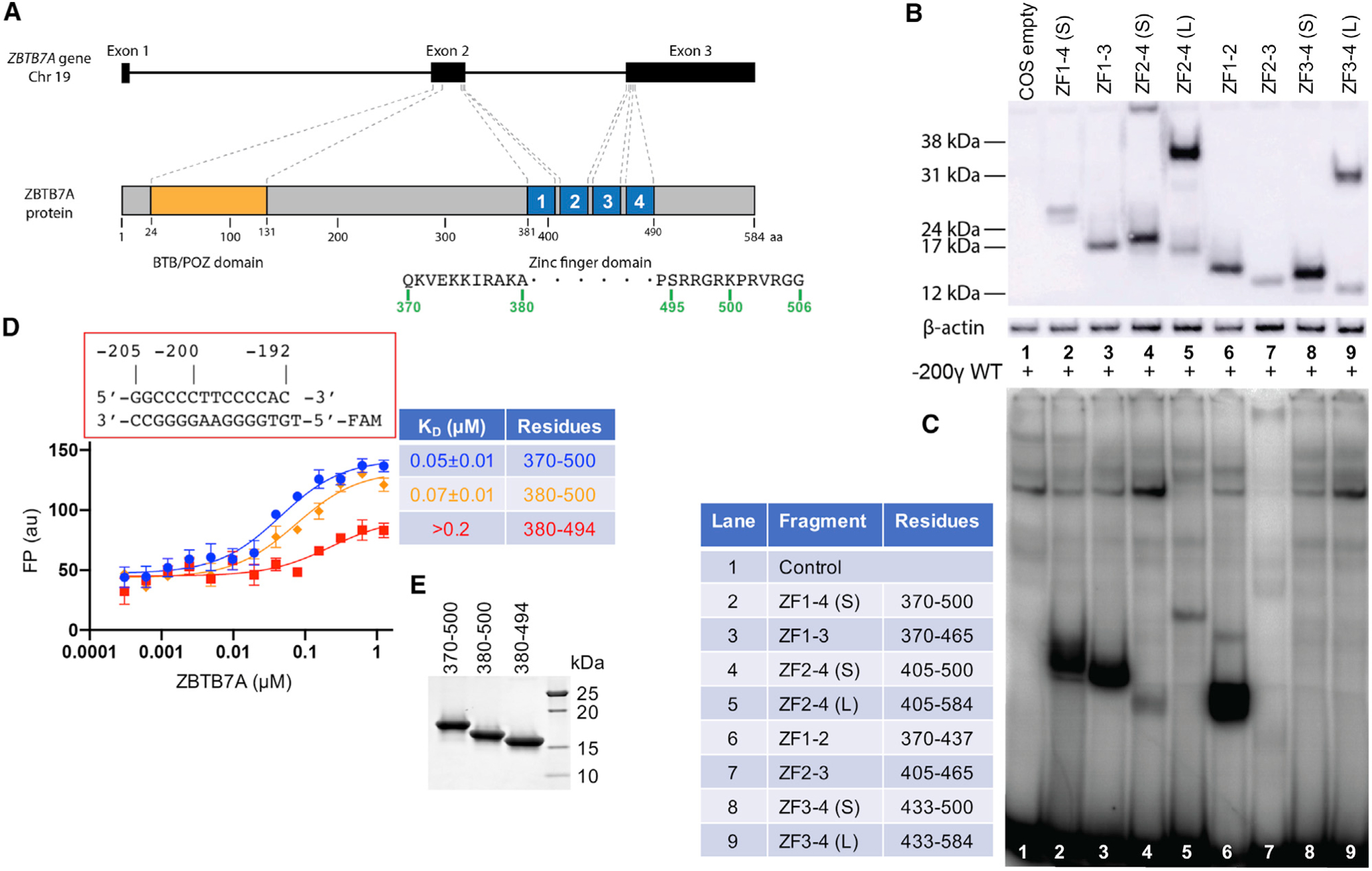
Figure 1. ZBTB7A DNA-binding domain.

(A) Schematic diagram of ZBTB7A gene and protein.
(B) A western blot of the nuclear extracts prepared from COS-7 cells transiently expressing constructs containing different numbers of ZFs of ZBTB7A using an antibody against the FLAG tag.
(C) An EMSA assay of the nuclear extracts assessing binding to a radiolabeled DNA probe covering the −200 element (−200γ wild type [WT]).
(D) FP assay of ZBTB7A ZF fragments expressed in E. coli with and without N- and C-terminal additions. Data represent the mean ± SD of N number of independent determinations (N = 3). For the percentage of saturation, see Figure S2.
(E) A 4%–20% gradient gel with Coomassie blue staining showing the examples of purified recombinant proteins used in the FP assays.
