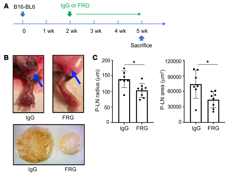
Figure 3. CHI3L1 regulates local footpad lymphatic spread of B16-BL6 cells.
B16-BL6 cells (2 × 10⁴) were injected into the right footpad. Two weeks later the mice were randomized to FRG or isotype control antibodies. After an additional 3 weeks the footpad lesions and popliteal lymph nodes were compared. (A) Treatment scheme used in these experiments. (B) Representative photographs of popliteal lymph nodes. The top panels compare the popliteal regions (indicated by arrows) of mice treated with control IgG versus FRG. The bottom panel compares popliteal lymph nodes from the right foot of mice treated with control IgG versus FRG. (C) Size (diameter and surface area) quantification of popliteal lymph nodes (P-LN). The values in C represent the mean ± SEM of the noted evaluations represented by the individual dots. *P < 0.05 (Student’s t test).

