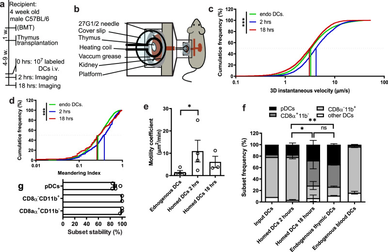
Fig. 2. Analysis of dendritic cells in endogenous and transplanted thymi.
a Experimental protocol for thymic MP-IVM experiments. BMT: bone marrow transplantation. b Schematic drawing of the experimental setup to image transplanted thymi by MP-IVM. c 3D instantaneous velocity and d meandering index of thymus-resident DCs and homed DCs at 2 hours and 18 hours post transfer. n = 3 independent experiments. e Motility coefficients of endogenous and homed DCs. n = 4 independent experiments for 2 hrs and n = 3 independent experiments for 18 hrs time points. f Subset composition of resident thymic DCs and homed DCs 2 and 18 h post transfer. n = 2 experiments with 10 mice/group. g FACS-sorted splenic DC subsets were fluorescently labeled and injected IV into recipient mice. 18 h later, subset-specific markers on homed DCs in recipient thymi were analyzed by flow cytometry. DC subsets were sorted as follows: pDCs: CD11c+ PDCA1+ B220+; CD8α+CD11b– DCs: CD11chigh PDCA1– B220– CD8α+ CD11b–; CD8α–CD11b+ DCs: CD11chigh PDCA1– B220– CD8α+ CD11b–. n = 5 mice/group. Bars and error bars represent mean ± SEM. Statistical comparisons were performed using two-way ANOVA unless stated otherwise. *P = 0.0428 (e) and P = 0.0142 (f); **P = 0.0026, ***P < 0.001; n.s. not significant. DCs dendritic cells, pDCs plasmacytoid dendritic cells. See also Supplementary Fig. 1 and Supplementary Movies 1 and 2.

