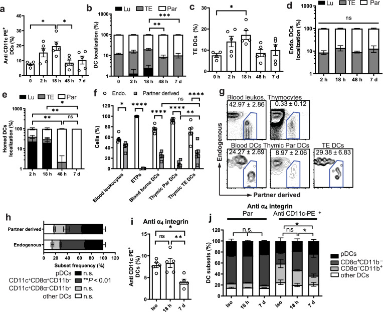
Fig. 5. Origin of thymic TE-DCs.
a–e C57BL6 mice (CD45.1+) were injected with congenic (CD45.2+) splenic DCs and analyzed at indicated time points. Two minutes prior to tissue harvest, mice received anti-CD11c-PE mAb IV (n = 3 experiments with five mice/group). Labeling indices were determined to identify DC localization as illustrated in Supplementary Fig. 3 and Fig. 4a. a Frequency of PE+ DCs among total DCs recovered from thymi before and after adoptive DC transfer (one-way ANOVA). b Frequency of TE-DC, Lu-DC, and Par-DC among total thymic DCs (two-way ANOVA) and c frequency of TE-DCs at indicated time points before and after DC transfer (one-way ANOVA). d Localization of endogenous DCs and e homed DCs at the indicated time points (two-way ANOVA). f–h Parabiotic pairs were generated by surgically joining age- and sex-matched CD45.1+ and CD45.2+ congenic mice. Two weeks after parabiosis surgery, animals were injected with anti-CD11c-PE and tissues were harvested 2 mins later. n = 2 experiments with five mice/group (two-way ANOVA). f Frequency of endogenous (empty bars) and partner-derived (filled bars) leukocytes, early thymic progenitors (ETPs), and DCs in blood and thymus. g Representative FACS plots of leukocytes and DCs in parabiotic animals. h Subset composition of endogenous and partner-derived thymic DCs. i and j Effect of α4 integrin inhibition on thymic DC subsets. C57Bl/6 mice were either treated for up to 7 days with a blocking anti-α4 integrin mAb or isotype control (Iso). Mice received anti-CD11c-PE mAb 2 mins prior to tissue harvest (n = 2 experiments with five mice/group; two-way ANOVA). i Frequency of CD11c-PE+ cells among total thymic DCs (one-way ANOVA). j Subset composition of thymic Par-DC and anti-CD11c-PE+ DCs (two-way ANOVA). Bars and error bars represent mean ± SEM. a *P = 0.0236 (0 h vs. 18 h) and P = 0.0302 (18 h vs. 48 h). b ***P = 0.0009, **P = 0.0068. c *P = 0.0405. e **P = 0.005 (2 h vs. 48 h) and P = 0.005 (2 h vs. 7d), *P = 0.0488 (18 h vs. 7d). f **P = 0.0019, *P = 0.041, ****P < 0.0001. i **P = 0.005, *P = 0.0115. j *P < 0.05; n.s. not significant, Lu luminal dendritic cells, TE transendothelial dendritic cells, Par parenchymal denritic cells, pDCs plasmacytoid dendritic cells. See also Supplementary Fig. 5.

